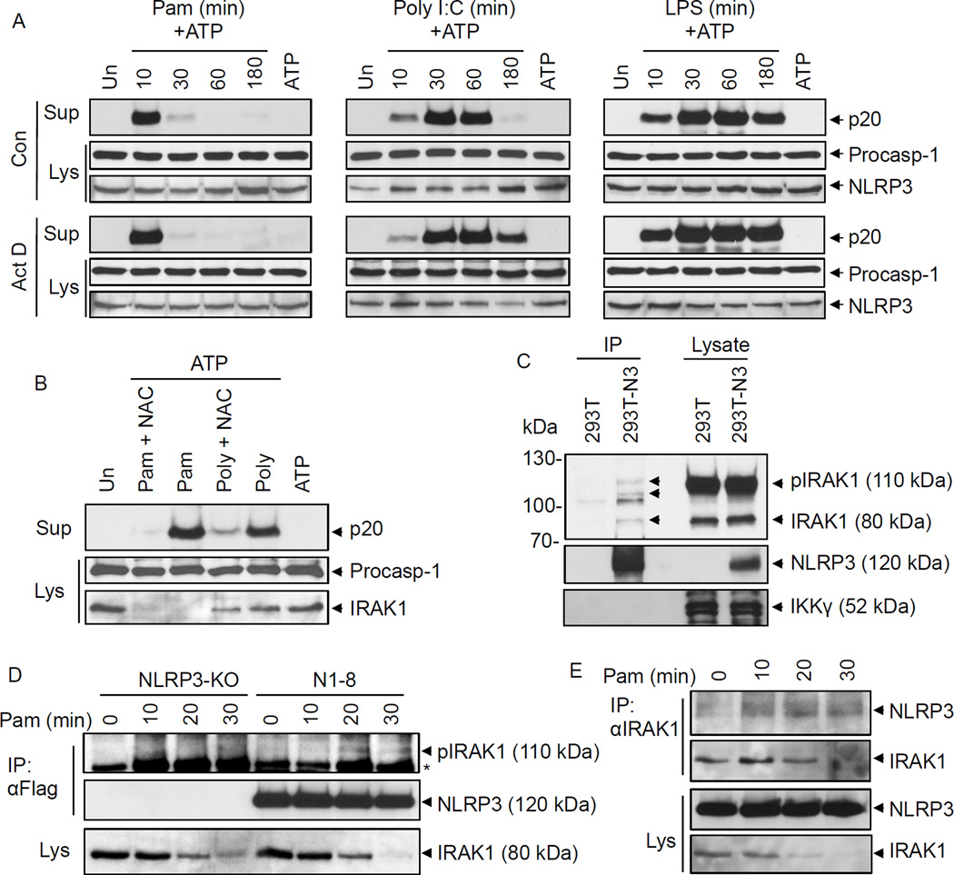
Figure 3. Transcription-independent ROS-dependent priming of the NLRP3 inflammasome.
A, B, immunoblots of caspase-1 in the culture supernatants (Sup) or cell lysates (Lys) of mouse WT macrophages primed with Pam3CSK4 (Pam), Poly I:C or LPS for the indicated times (min) followed by stimulation with ATP for 45 min in the absence (Con) or presence of actinomycin D (Act D, 0.5 µg/ml) (A), or primed with Pam3CSK4 (Pam for 10 min) or poly I:C (Poly for 45 min) followed by stimulation with ATP for 45 min in the absence or presence of N-acetyl cysteine (NAC, 25 µM) (B) as indicated. The bottom panels show NLRP3 (A) or IRAK1 (B) in the same cell lysates. C, immunoblots showing NLRP3 associates with IRAK1 when NLRP3 was immunoprecipitated (IP) from 293T-NLRP3 cells (293T-N3) but not 293T cells. 293T and 293T-N3 were transfected with WT IRAK1 plasmid. The bands marked with arrows indicate interacting IRAK1 isoforms. The bottom panel shows IKKγ as a negative control. D, E, immunoblots showing NLRP3 associates with endogenous IRAK1 when NLRP3 (D) or IRAK1 (E) were immunoprecipitated (IP) from macrophages following stimulation with Pam3CSK4 for the indicated times. N1–8 represents macrophages stably reconstituted with Flag-NLRP3. NLRP3-KO cells were used as control. Asterisk indicates a non-specific band.

