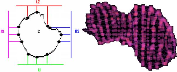
Figure 4.

Parameterization points. Left - Uniformly spaced points are projected onto the object in the convexity plane shown in different colors. Right – the parameterization points are highlighted as small cubes on the surface of a liver with irregular shape, when 13 convexity planes were used. These points allow point-to-point correspondence between two shapes.
