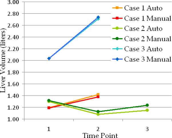
Figure 8.

Examples of change over time in liver volume for three cases from the clinical data with small (case 1- red), moderate (case 2- green) and large (case 3- blue) metastatic liver tumors. Manual and automated estimations are presented for comparison. Image data for cases 1 and 3 are shown in Figure 7.a and 7.b respectively.
