
Figure 6.
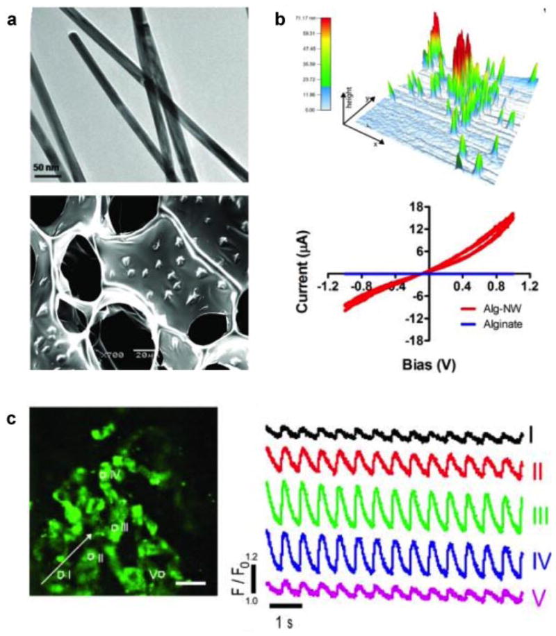
Nanocomposite hydrogels containing gold nanowire and alginate. a: The distribution of gold nanowires determined using TEM indicates an average length of ~1 μm and an average diameter of 30 nm. The uniform distribution of nanowire within the alginate scaffolds was also observed. b: The topographic mapping of nanocomposite detects the presence of nanowires. In nanocomposite, the current increased with bias voltage over the range −1 to 1V, while negligible current was passed through alginate (without nanowire) over that same range. c: Calcium transient was determined at various locations using calcium dye. The number indicates location of on the Alg-gold nanowire scaffold and the corresponding curves indicate calcium transport at those points. Adapted with permission from Dvir et al. (2011). Copyright (2013) Nature.