
Figure 3.
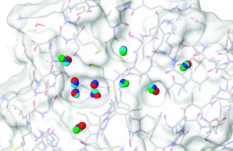
The molecular surface of the MAYM mutant showing the positions of ten water molecules in the crystal structure and the predicted hydration sites from the three simulation schemes. The oxygen atoms of the crystal structure water molecules are colored red, the hydration sites from the free simulation are colored green, the hydration sites from the restrained simulation are colored dark blue, and the hydration sites from the fixed simulation are colored cyan.