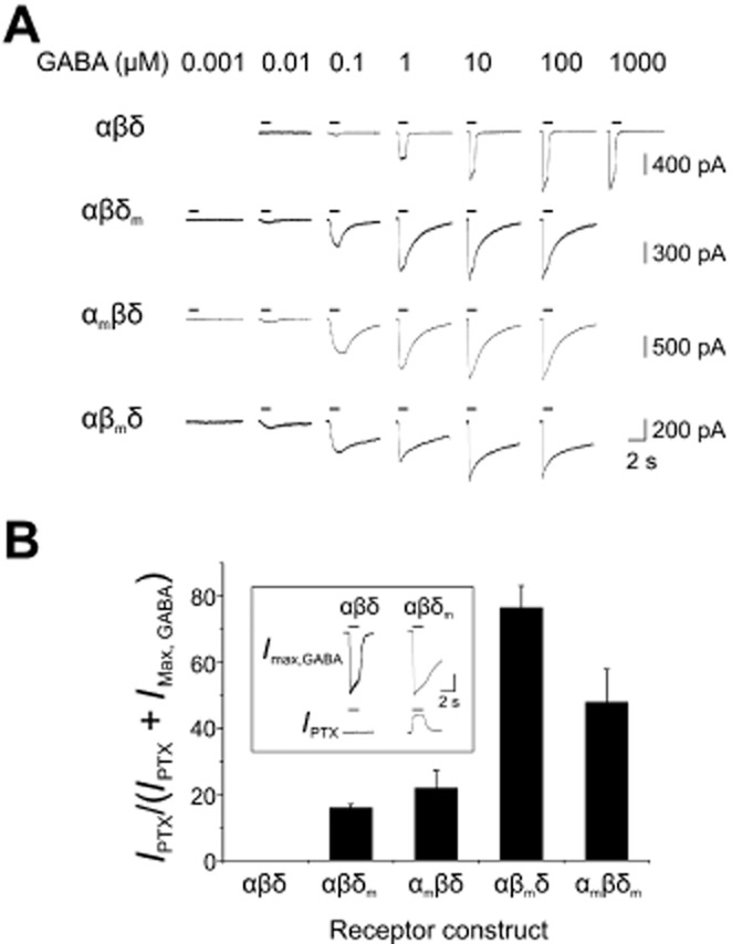
Figure 2.

Functional expression of WT and L9′S mutant α4, β3 and δ subunits. In this and subsequent figures, WT α4, β3 and δ subunits are labelled as α, β and δ, whereas mutated α4(L297S), β3(L284S) and δ(L288S) subunits are designated as αm, βm and δm. (A) Examples of whole-cell currents elicited by increasing concentrations of GABA on HEK cells expressing recombinant αβδ, αβδm, αmβδ and αβmδ receptors. A transfection ratio of 10α:1β:10δ was used. Note the increased GABA sensitivity and prolonged deactivation kinetics exhibited by mutant-expressing cells. (B) Bar graph of SA for αβδ, αβδm, αmβδ, αβmδ and αmβδm receptors. Values were calculated by expressing the outward current induced by the Cl− channel blocker picrotoxin (IPTX; 1 mM) as a percentage of the maximum current, defined as the sum of IMax,GABA and IPTX (n = 4–11; mean ± SEM). No SA (= 0%) was observed for WT α4β3δ receptors. The inset shows example GABA-activated and picrotoxin-sensitive currents (IMax,GABA and IPTX) for αβδ and αβδm receptors. Current calibration bars: 300 pA (αβδ); 400 pA (αβδm).
