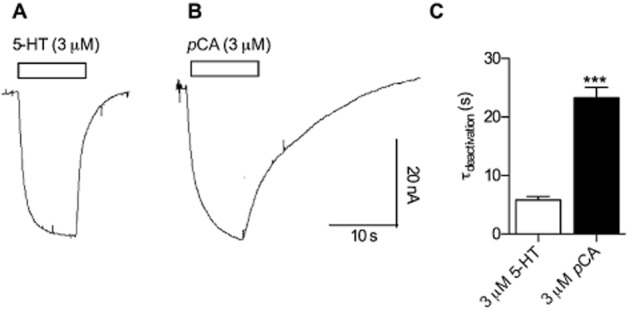
Figure 1.

Differences between currents induced by 5-HT and pCA respectively. Xenopus laevis oocytes expressing hSERT were clamped to −60 mV. Currents induced by 3 μM 5-HT were compared with currents from the same cell induced by 3 μM pCA. (A) 3 μM 5-HT was applied to the cell for 10 s and subsequently washed away with a gravity driven perfusion system. (B) The same procedure was repeated with 3 μM pCA. (C) The current decays were fitted to mono-exponential functions and the time constants were plotted in the bar graph. A comparison of the decay time constants following 5-HT (5.8 s ± 1.3 s, n = 6) and pCA removal (23.3 s ± 4 s, n = 6) revealed a significantly slower time constant for pCA (*** = P < 0.001, Mann–Whitney U-test).
