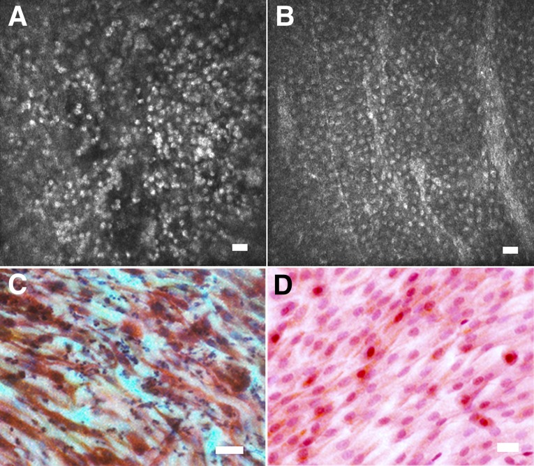
Figure 5.
Nonstratifying epithelial phenotype observed in in vivo confocal microscopy and impression cytology observed in several patients with limbal stem cell deficiency. Shown are confocal microscopy (A, B) and corresponding impression cytology (C, D) images from the central cornea of two patients with limbal stem cell deficiency prior to treatment. (A, B): Confocal microscopy shows a thin sheet of small cells, usually a mono- or bilayer, with hyperfluorescent nuclei, but no other cellular detail is visible. (C, D): Correlation with impression cytology performed on the same patients directly after confocal scans showed that these cells had a spindle-like shape and expressed CK19 (red) but not CK3 (brown) (C, D). This phenotype may represent conjunctival cell migration onto the corneal stroma but failure of the cell layer to form cell junctions and differentiate into a mature multilayered epithelium. Scale bars = 25 μm.

