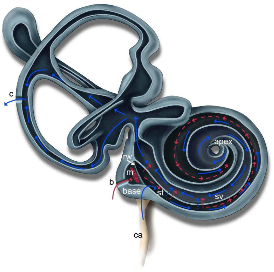
Figure 1.
Illustration of the mouse inner ear depicting the hypothesized advection flow induced by creation of a fluidic exit via canalostomy (c) in the posterior semicircular canal. Drug is delivered to the round window (rw) via polyimide microtubing inserted through a bullaostomy (b) into the middle ear (m) and positioned at the RWM niche. The drug is absorbed through the RWM into scala tympani (st). Diffusion is shown in red as dotted lines. Induced advection flow due to CSF influx from the cochlear aqueduct (ca) is shown as solid lines in blue. This flow carries the drug throughout the cochlea via a flow path through st, to the helicotrema at the apex, through scala vestibuli (sv) and into the vestibular system to the canalostomy (c). Adapted from [14].

