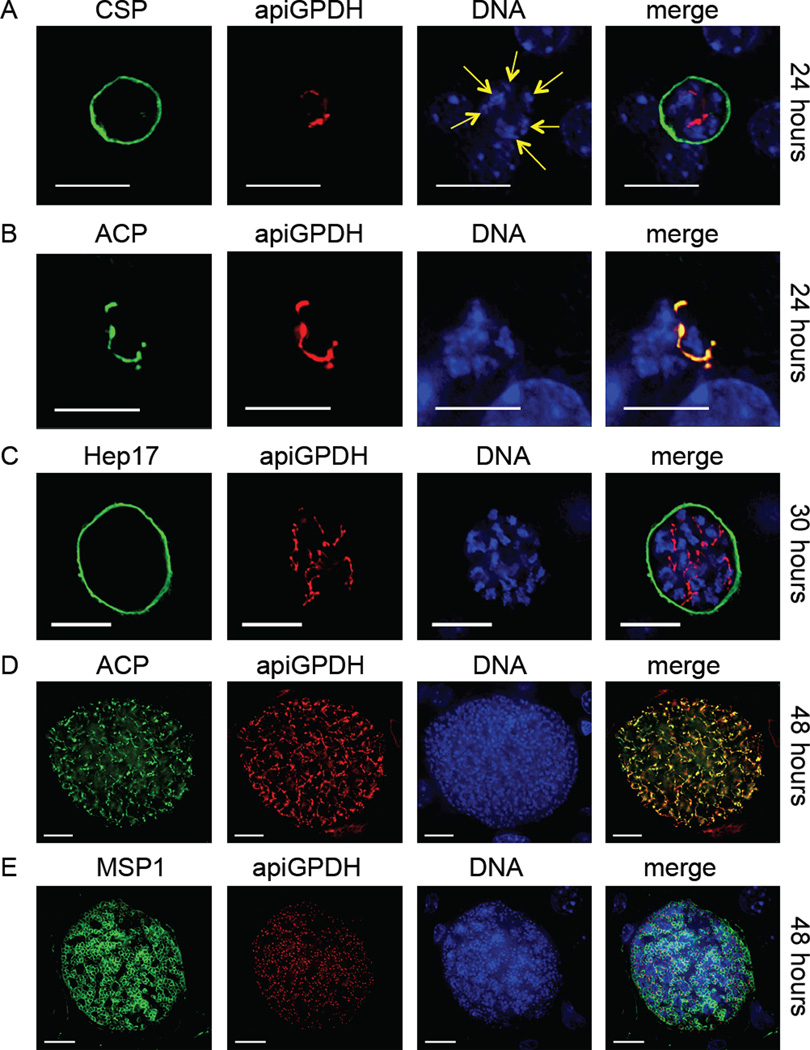
Figure 1.
The Plasmodium glycerol 3-phosphate dehydrogenase (apiG3PDH) predicted to target to the apicoplast is expressed only during liver stages and co-localizes with the apicoplast lumen-targeted acyl carrier protein (ACP). The expression of apiG3PDH was assessed with the transgenic epitope-tagged Py apiG3PDHmyc parasite and identified by IFA. At 24 hours (A), nuclear replication was apparent (yellow arrows point to multiple nuclei centers) and apiG3PDH expression was in internal structures reminiscent of the apicoplast. Antibody to circumsporozoite protein (CSP) was used to delineate the parasite plasma membrane (PPM). It was also apparent at 24 hours (B), that apiG3PDH perfectly co-localized with the apicoplast lumen protein acyl carrier protein (ACP). At 30 hours (C), liver stages had increased in size, as had the complexity of apiG3PDH localization, which was contained within the parasitophorous vacuole membrane (PVM) marker Hep17. At 48 hours (D), co-localization of apiG3PDH with ACP clearly shows that apiG3PDH is localized to the extensively branched apicoplast in a late-liver stage parasite. Fully mature liver stages at 48 hours showed the presence of merozoites, localized with the PPM marker merozoite surface protein 1 (MSP1) and each merozoite, as expected, appeared to contain a single apicoplast, based on apiG3PDH expression (E). DNA was stained with 4',6-diamidino-2-phenylindole. Scale bar: 10 µm.

