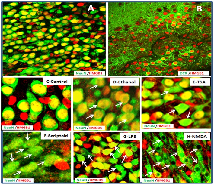
Figure 2. Neuronal expression of HMGB1.
Representative confocal merger images showing A: double immunofluorescent staining with anti-HMGB1 (red) and anti-NeuN (green), a marker for mature neurons. Majority of NeuN+ neurons coexpressing HMGB1 (yellow); B: double immunofluorescent staining with anti-HMGB1 (red) and anti-doubleCortin (DCX, green), a marker for immature neurons. HMGB1 is located in the nuclear of majority DCX+ neurons. Mobilization and translocation of nuclear HMGB1 in HEC slices was further depicted from treatment of Control (C), ethanol (D), TSA (E), Scriptaid (F), LPS (G) and NMDA (H). Nuclear mobilization and/or cytoplasm translocation in neurons were indicated by arrows (original magnification 80x in A–B: 320x in C–H).

