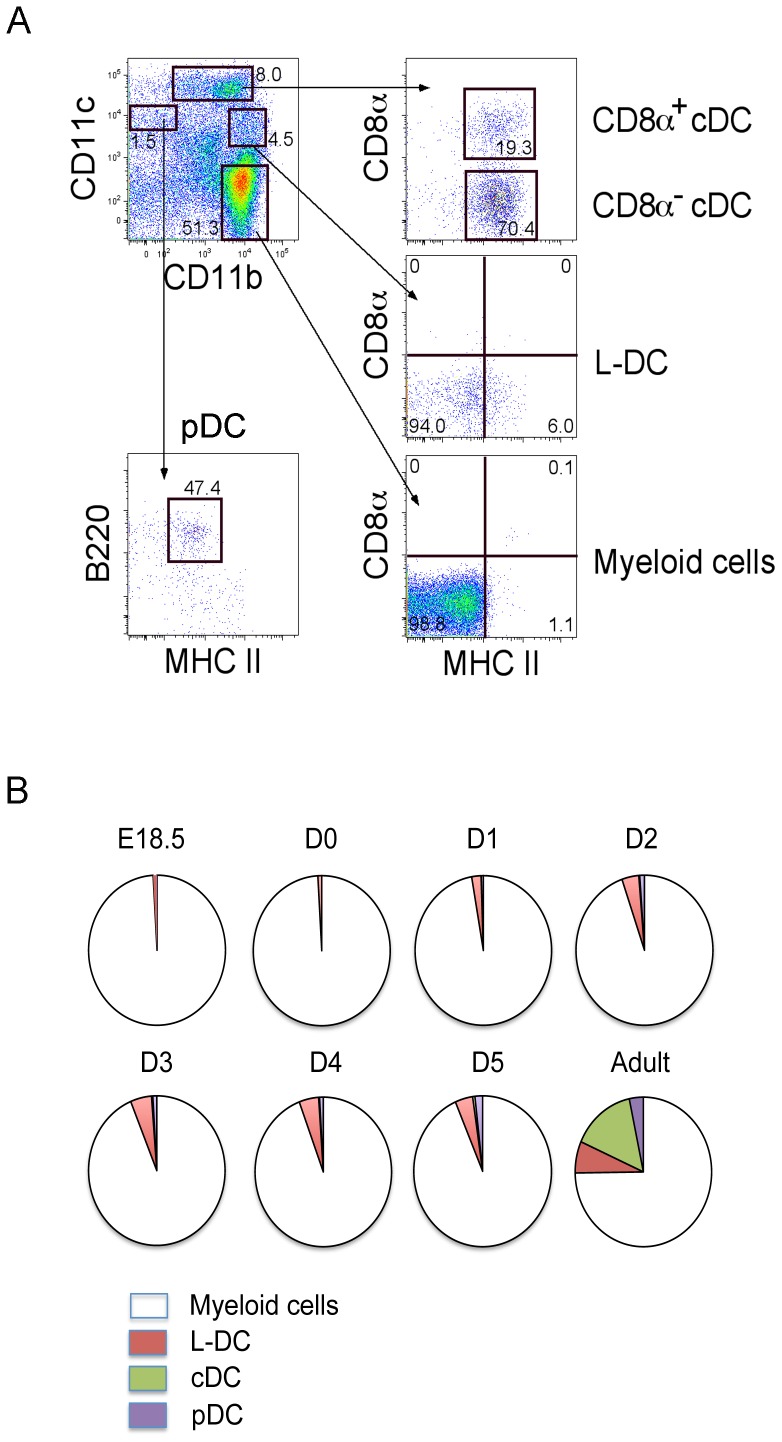
Figure 1. Characterization of splenic DC subsets.
(A) Perinatal and adult spleen cells from C5BL/6J mice were stained with antibodies to distinguish DC subsets flow cytometrically. The procedure for gating subsets in one adult mouse is shown by example. Propidium iodide (PI) staining was used to discriminate dead cells. Gates were set on bivariate plots using isotype control antibodies and numbers on gates reflect % positive cells. CD11b and CD11c staining was used to identify CD11bhiCD11c−, CD11bhiCD11clo, CD11b+CD11chi and CD11b−CD11c+ subsets. Further staining for CD8, MHC-II and B220 was used to distinguish CD8+ cDC and CD8− cDC, L-DC, myeloid cells, and pDC. (B) The proportional representation of myeloid and DC subsets amongst the total CD11b+ and/or CD11c+ dendritic/myeloid population is shown. Data were derived using average values obtained for 2 mice. E = embryonic; D = day post birth.

