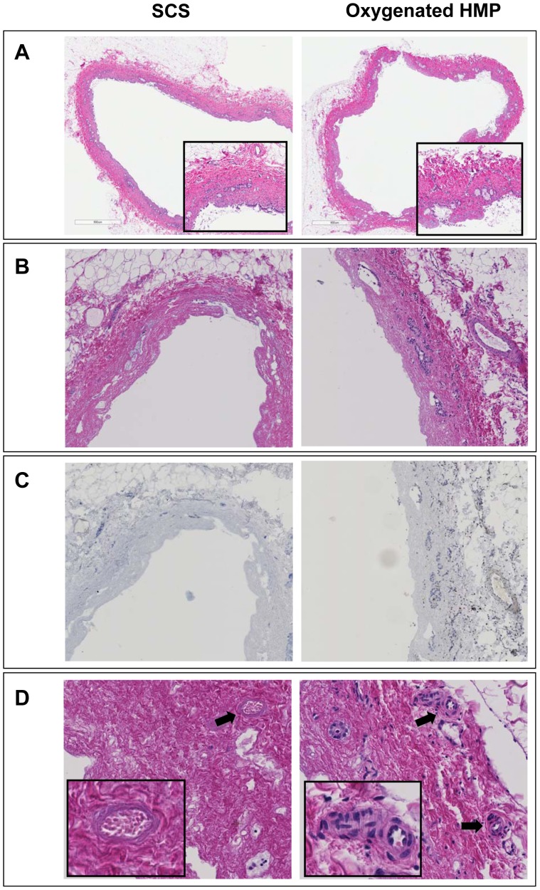
Figure 6. Representative examples of H&E histology of bile ducts of DCD liver grafts preserved by either 4 h of oxygenated HMP or 4 h of SCS followed by 2 h by normothermic ex vivo sanguineous perfusion.
Panel A: extrahepatic bile duct immediately after procurement (magnification 200x). The insert represents a higher magnification of 400x. Panel B: extrahepatic bile duct after preservation and reperfusion (magnification 280x). Panel C: Immunohistochemistry for activated caspase-3 of the same bile ducts as presented in panel B (brown color indicates immunopositivity; counterstaining with hematoxylin). Very few caspase-3 positive cells were detected in the bile duct wall stroma. Remnant biliary epithelial cells (i.e. in the peribiliary glands) were not positive for acitivated caspase-3. Panel D: higher magnification (400x) of extrahepatic bile ducts focusing on the peribiliary plexus. Arrows indicate peribiliary arterioles. The insert represents a higher magnification of the peribiliary arterioles (800x). Bile ducts of livers preserved by oxygenated HMP displayed significantly less signs of arteriolonecrosis, compared to livers preserved by SCS (see also Table 1 ).

