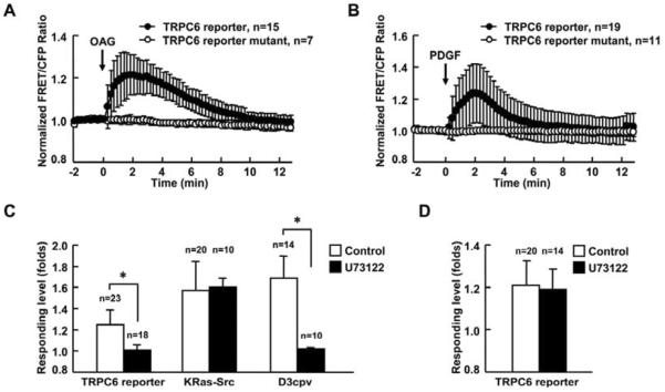
Fig. 2. PDGF activates TRPC6 via the PLC pathway in MEFs.
(A) and (B) Representative time courses of normalized FRET/CFP emission ratio of TRPC6 reporter (black dots) and its mutant (white dots) upon (A) 300 μM OAG or (B) 10 ng/ml PDGF stimulation. (C) Responses of TRPC6, KRas-Src or D3cpv reporter to 10 ng/ml PDGF stimulation in MEFs with (solid bars) or without (open bars) 2 μM U73122 pretreatment for 5 min. MEFs transfected with the TRPC6 reporter were pretreated with 1 μM TG for 1 hr before imaging to deplete the calcium storage ofintracellular organelles. MEFs transfected with D3cpv were maintained in Ca2+ free HBSS buffer during imaging to eliminate the calcium influx across the plasma membrane. (D) Responses of TRPC6 reporter to 300 μM OAG stimulation in MEFs with or without 2μM U73122 pretreatment for 5 min. The data represents the means±SD from multi-samples. “n” represents the cell number in each group. * indicates the significant difference between indicated groups (P < 0.01).

