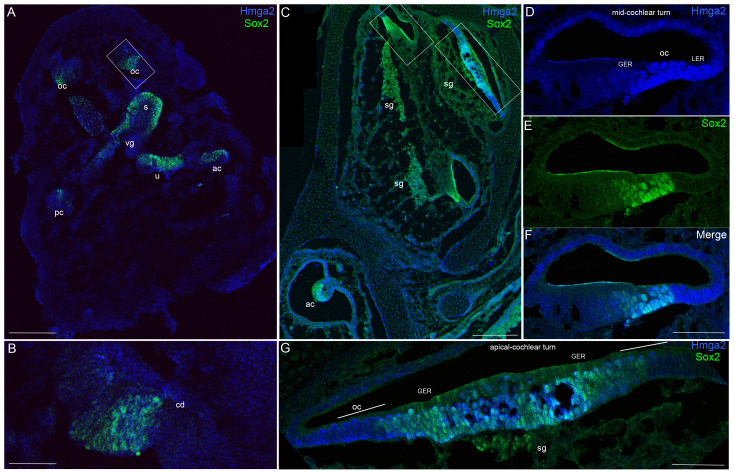
Figure 2. Hmga2 expression pattern in the embryonic E13 (A–B) and E14.5 (C–G) mouse inner ears.
Low-magnification micrograph of inner ear longitudinal section immunostained with Hmga2 (shown in blue) and Sox2 (shown in green) antibodies (A). In the E13 inner ear longitudinal section, a vestibular ganglion in the boundary of the saccule have a relatively weaker Hmga2 expression, while no obvious Hmga2 signal at other regions of the inner ear. In contrast, Sox2 expression marked the prosensory domains of the cristae (ac, pc), utricular macula (u), saccular macula (s) and the organ of Corti (oc). Higher-magnification view of the inner ear longitudinal section (B) at the level of the region indicated by the box in (A). In the developing cochlear duct (cd), Sox2 marked the prosensory region of the organ of Corti. No obvious signal for Hmga2 expression was detected in this Sox2 prosensory region. Low-magnification micrograph of E14.5 inner ear longitudinal section immunostained with Hmga2 (shown in blue) and Sox2 (shown in green) antibodies (C). (D–F) Higher-magnification view of the inner ear longitudinal section at the level of the mid-cochlear turn in the region indicated by the small box in (C). Hmga2 expression is observed in the nuclei of hair cells and supporting cells of the organ of Corti's and in the GER and LER cells. In the OC, Hmga2 (D,F, blue) co-localized with Sox2 (E,F, green) in the hair cells and supporting cells. (G) Higher-magnification view of the inner ear longitudinal section through the apical cochlear turn in the region indicated by the large box in (C). Hmga2 immunolabeling is localized to lateral halves of the cochlear duct (the site of the future OC) and was present in the GER throughout the thickness of the epithelium (shown in Blue) where it is colocalized with Sox2 (shown in green). Organ of Corti (OC), spiral ganglion (sg); vestibular ganglion (vg); cochlear duct (cd); lesser epithelial ridge (LER); greater epithelial ridge (GER), anterior crista (ac); posterior crista (pc). Scale bars = 200 µm in (A,C) and 50 µm in (B,D,E,F,G).

