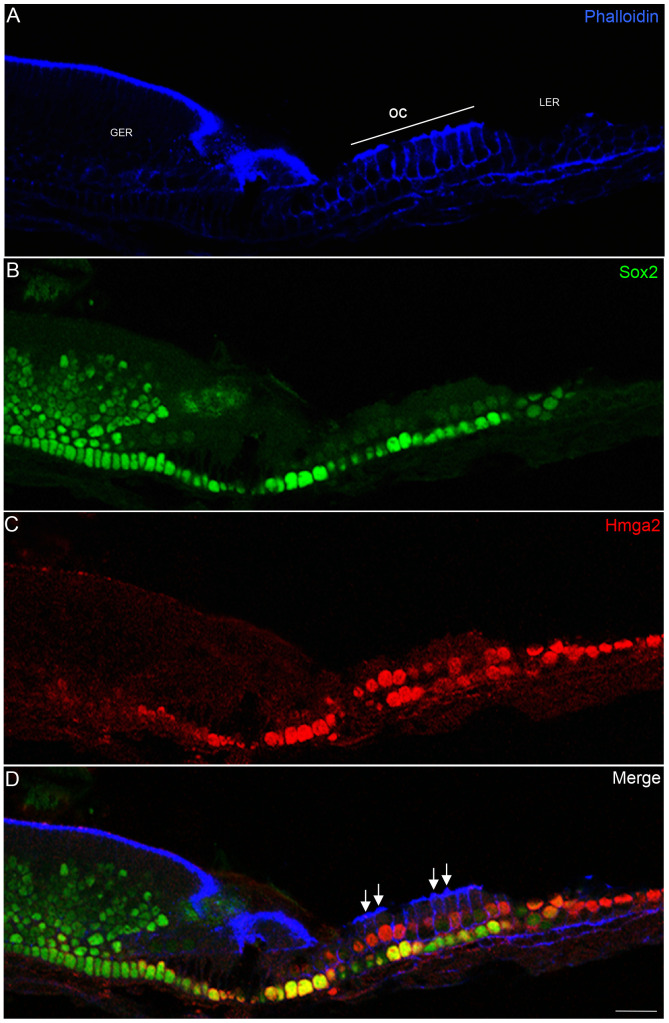
Figure 4. Hmga2 immunostaining in the newborn cochlea.
Tangential section through the mid-turn (z-section) of the cochlea showing widespread expression of Hmga2 (shown in red) in the nuclei of hair cells and supporting cells; in the LER region; Deiter's cells and in pillar cells. In the organ of Corti area, Hmga2 is colocalized (shown in yellow) with Sox2 in the supporting cells. Phalloidin marker (shown in blue) was used to outline the general structure of the cochlear epithelium including stereocilia (D, arrows) and the cellular borders of hair and supporting cells. Scale bars = 20 µm in all panels.

