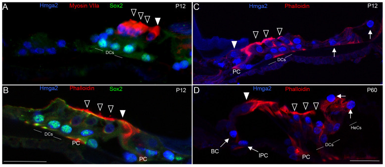
Figure 6. Hmga2 expression is maintained with Sox2 in postnatal day-12 (P12) and in adult (P60) cochleas.
By P12 (A–C), Hmga2 expression (shown in blue) is still observed in the nuclei of both hair cells co-labeled with the Myosin VIIa (shown in red, A) and supporting cells co-labeled with Sox2 (shown in green, A–B). A transverse section of P12 organ of Corti's in mid-cochlear turn (C) co-labeled with Hmga2 (shown in blue) and phalloidin (shown in red) confirming the expression of Hmga2 in the hair and supporting cells. In the adult (D), Hmga2 immunolabeling (shown in blue) is maintained in the nuclei of hair and supporting cells of the organ of Corti. The supporting cells immunolabeled with Hmga2 included the Deiters cells (DCs), Pillar cells (PCs), inner phalengeal cells (IPC), Border cells (BC) and Hensen cells (HeCs, arrows). The organ of Corti outline is visualized by phalloidin-labeling (red color, B–D). Open arrowheads indicate the OHCs and filled arrowhead indicates the IHC (A–D). Scale bars = 20 µm in (A) all panels.

