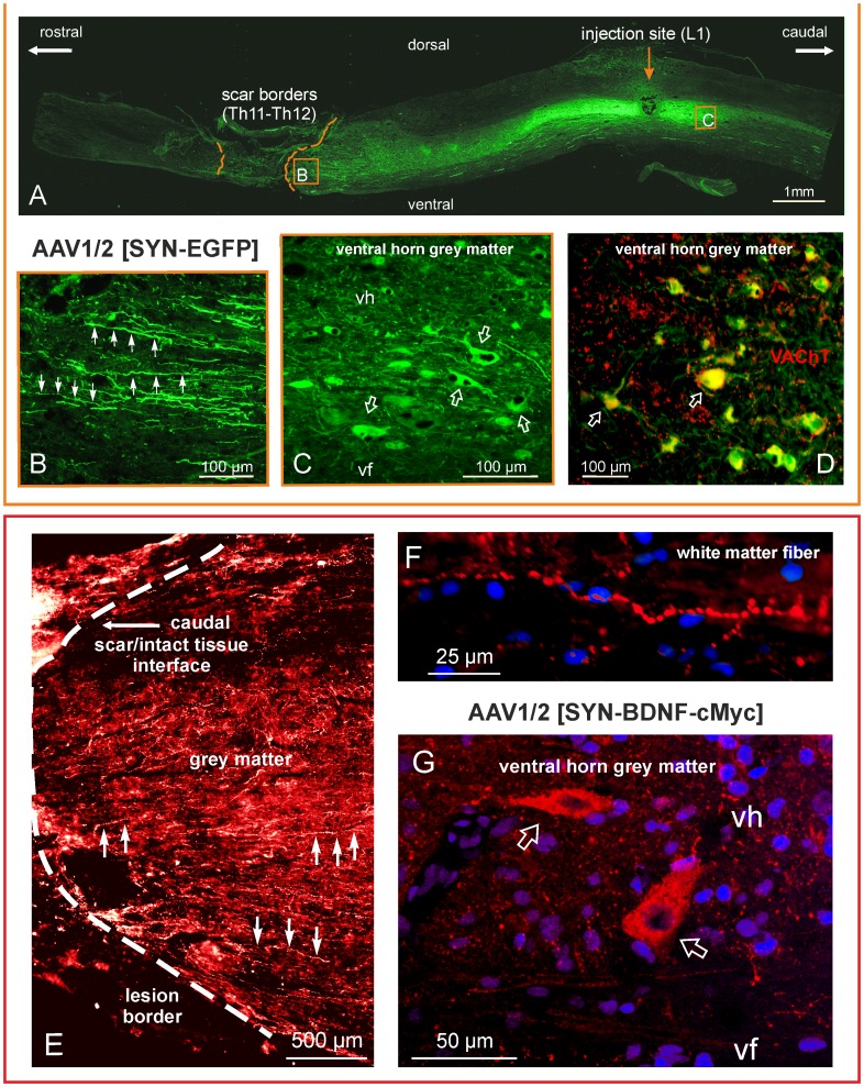
Figure 1. Spinal cord transduction with an AAV1/2 vector expressing EGFP or BDNF with cMYC tag at 7 weeks after spinal cord transection.
Upper panel: (A) Tiled images taken of the thoraco-lumbar segments of a spinal cord from a spinalized rat that received the EGFP transgene; micrographs were taken on a fluorescence microscope at ×10 magnification. The dashed lines delineate the edges of the scar. (B) An enlarged micrograph of the framed area in (A) documenting the numerous EGFP-positive fibers (arrows) running along the grey matter in close proximity to the lesion site. (C) An enlarged micrograph of the framed area in (A) showing transduced cells of neuronal morphology in the ventral horn (arrows). (D) A merging of the EGFP expression signal (green) with vesicular acetylcholine transporter (VAChT, red) immunolabeling shows their colocalization (yellow), which confirms that the transduced cells are motoneurons (arrows). Bottom panel: The spinal cord from a rat that received the BDNF-cMYC transgene. (E) cMYC immunostaining detected BDNF-cMYC-positive neuronal fibers (exemplified by arrows) below the transection, in the lower thoracic segments of the spinal cord. Fibers approach and encroach on the scar from its caudal aspect. The white dashed line delineates the caudal border of the scar. A dense mesh of small caliber BDNF-cMYC-positive fibers prevails in the grey matter (E) whereas large caliber varicose fibers appear in the white matter (F). (G) The confocal microscope microphotograph shows two BDNF-cMYC-positive, large size neurons (arrows) of the L2 ventral horn. Hoechst nuclear labeling is shown in blue. Abbreviations: vh – ventral horn, vf – ventral funiculus.

