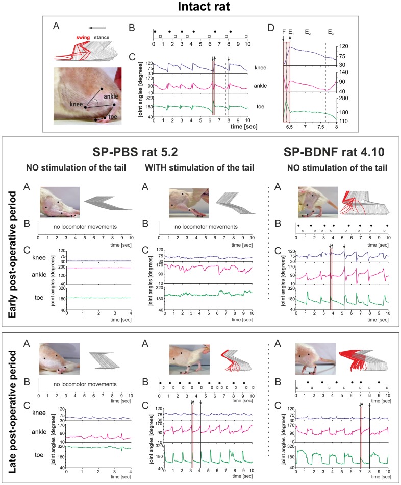
Figure 3. BDNF overexpression leads to an early recovery in locomotor function. Comparison of the treadmill locomotion of the intact, SP-PBS and SP-BDNF rats.
Upper panel: kinematic analysis of the gait of an intact rat during locomotion on the moving treadmill (A) Stick figure showing the angular excursions of the knee, ankle and toe joints during one step cycle during slow (0.05 m/s) treadmill locomotion in an intact rat. To measure the angular excursion of the hindlimb joints, the black markers were glued to the shaved skin overlying the femur and tibia heads, tibiofibular articulation, distal metatarsus and distal phalanx of the third toe (bottom, A). A digital Panasonic camera (NV-GS400 3CCD) was used to capture video images of the hindlimbs during treadmill locomotion. Software based on Image–Pro Plus was used to create the stick figures of the hindlimb movements with a resolution twice as fast as that of the camera (i.e., 50 images/s). Every stick image was artificially separated from the next by the same coefficient to avoid superimposing neighboring stick images. (B) The footprints of both hindlimbs of an intact rat corresponding to the beginning of the foot contact with the treadmill during locomotion as taken from the video. Black dots - left leg; squares - right leg. (C) Angular excursions in the knee, ankle and toe joints during 10 s of treadmill locomotion. Downward deflection of the angular traces indicates flexion movement. (D) A framed plot from C of the angular excursions during one step was enlarged to indicate the phases of locomotion (F-E1 correspond to the swing and E2–E3 to the stance phase). Middle panel: examples of treadmill locomotion during the early post-operative period (the second week after surgery) of the spinal rats injected with PBS or with AAV-BDNF. None of the SP-PBS rats were able to perform locomotor movements when their hindlimbs were placed on the moving treadmill (exemplified in the left column; rat 5.2). Addition of tactile stimulation of the tail produced some agitation in both hindlimbs but did not evoke locomotor movements in the PBS-treated rats (central). Contrary to SP-PBS treated rats, no tail stimulation had to be used to trigger locomotion with body weight support in SP-BDNF rats. Periods of alternating, treadmill walking with body weight support and plantar foot placement but reduced rhythmicity were observed in eight out of the eleven rats overexpressing BDNF (exemplified in the right column, rat 4.10). Bottom panel: examples of treadmill locomotion of the spinal rats in the late (about 40 days) postoperative period. None of the SP-PBS rats could support their body weight or perform plantar foot placement (left); tail stimulation triggered locomotor movement in the PBS-treated rats (central). The locomotor capabilities of the hindlimbs in the SP-BDNF group improved profoundly in rats that were previously classified at the lowest level of the mBBB scale, whereas worsened in rats that walked well in the early post-surgery period (exemplified in the right column, rat 4.10). In that group, stimulation of the tail attenuated the quality of locomotion in rats that walked well without tail stimulation ( Table 2 , rats with scores 16–19). Weight support is defined as an elevation of the hindquarter [56].

