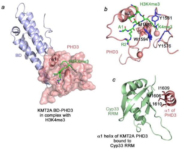
Figure 2. The structural basis of the interactions of KMT2A PHD3 with H3K4me3 and Cyp33.
(a) The 1.9 Å-resolution crystal structure of the PHD3-BD region of KMT2A in complex with histone H3K4me3 peptide (PDB: 3LQJ). (b) The H3K4me3-binding site of the KMT2A PHD3 finger. (c) The solution structure of the Cyp33 RRM domain fused with the α1-helix of the KMT2A PHD3 finger (PDB: 2KU7).

