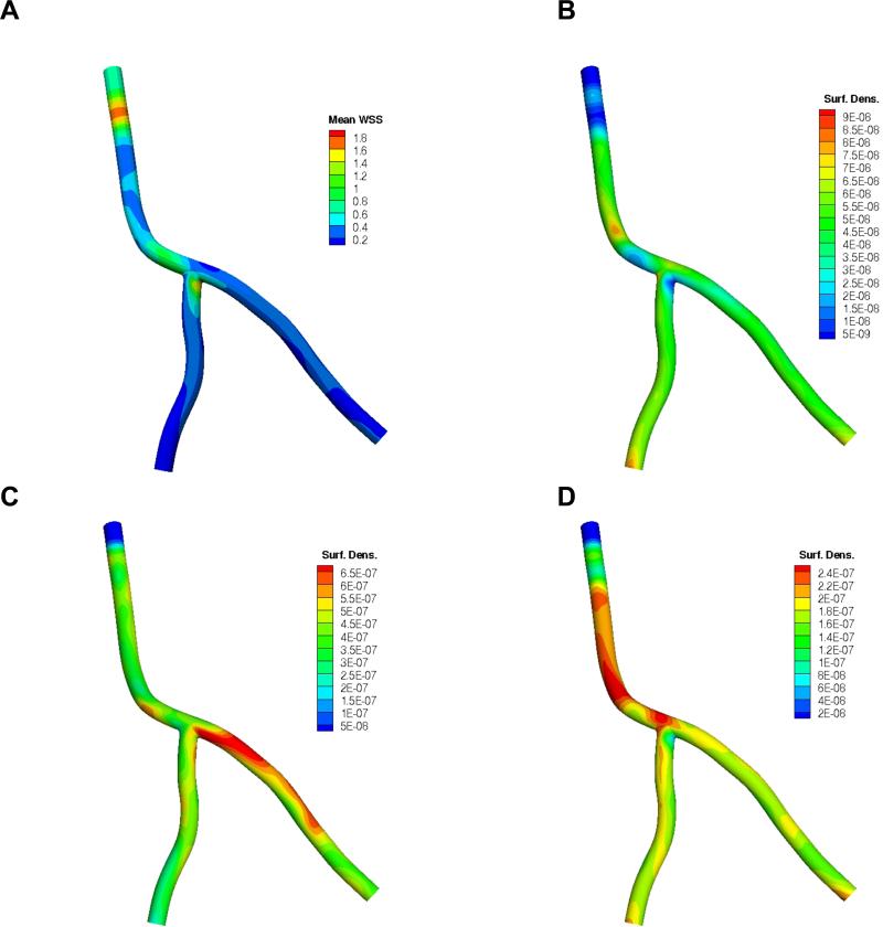
Figure 4.
A) Time-averaged wall shear stress (mean WSS) distribution in the coronary artery segment (with catheter at inlet) in Pa (N/m2); and the corresponding surface density (cm-2) of 0.5 μm particles at the end of simulation (t = 9 s) in terms of nadh /(ninj ×A) for the 3 targeted receptors: B) ICAM-1, C) VCAM-1, D) E-selectin. Note: color map scales are different. Here nadh is the number of adhered particles, ninj is the total number of injected particles and A (cm2) is the surface area.

