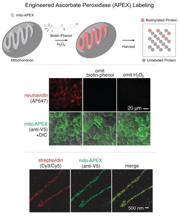
Figure 5.
(Top) Selective labeling of the mitochondrial matrix proteome in living cells requires 1) genetically targeting APEX to the mitochondrial matrix (mito-APEX), 2) initiating biotinylation by adding biotin-phenol and H2O2 to the medium, and 3) stopping biotinylation by cell fixation or lysis. (Middle) In human embryonic kidney cells, only mitochondria that expressed mito-APEX and were exposed to both biotin-phenol and H2O2 contained biotinylated proteins (stained with NeutrAvidin-Alexa Fluor 647). Both confocal fluorescence imaging (Middle) and stochastic optical reconstruction microscopy (STORM) (Bottom) showed that biotinylated proteins (stained with Streptavidin-Cy3/Cy5 for STORM images) overlapped with mito-APEX only in the mitochondrial matrix. (Adapted with permission from Rhee et al., Science, 339, 1328–1331, 2013. Copyright 2013 The American Association for the Advancement of Science.)

