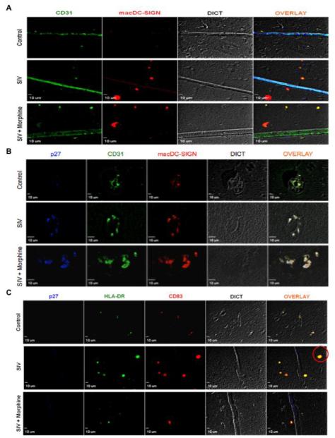
Figure 2. Dendritic cell infiltration into the CNS during SIV infection.
(A) After IHC-P staining, individual blood vessels in the frontal cortex were identified by CD31 staining (green) and macaque DC-SIGN staining (red) identified dendritic cells. The control group does not exhibit macDC-SIGN+ cells, however in SIV infection DCs are shown to transmigrate into the brain parenchyma. (B) Perivascular cuffing of dendritic cells is observed in SIV and SIV + morphine macaques at the site of SIV lesions as determined by staining with CD31 (green), macDC-SIGN (red) and SIV p27 (blue). Control animals do not show perivascular accumulation of lymphocytes. (C) Dendritic cells with a mature phenotype are observed in SIV and SIV + morphine animals, compared to control animals, as demonstrated by staining with HLA-DR (green) and CD83 (red) (red circle). Representative images at 40X magnification are shown.

