
Figure 5.
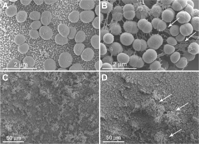
Staphylococcus epidermidis adhering to AuNP and Au after 24 and 48 hours, as visualized with SEM. After 24 hours, S. epidermidis on smooth gold surfaces (B) displayed more cell-connecting slime (some indicated by arrows) compared with nanostructured gold (A). A mature biofilm with high tower formations (arrows) was seen on smooth surfaces (D) after 48 hours, whereas the bacteria on nanostructured gold (C) were more horizontally scattered.
Abbreviations: AuNP, nanostructured gold surface; Au, smooth gold surface; SEM, scanning electron microscopy.