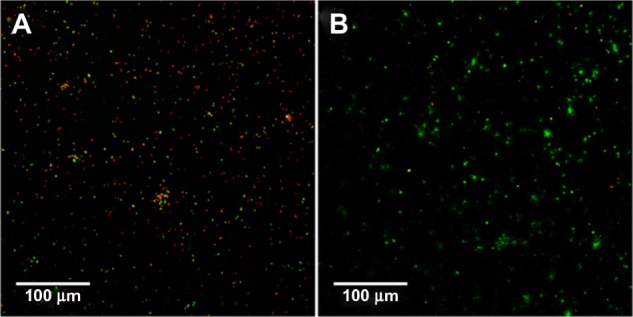
Figure 8.

Live and dead fluorescence staining of Staphylococcus epidermidis at the surface interface (z=0–3 μm) on AuNP (A) and Au (B) surfaces after 24 hours of static incubation in Roswell Park Memorial Institute medium, as analyzed by CLSM.
Note: Green indicates live cells; red indicates dead cells.
Abbreviations: Au, smooth gold surface; AuNP, nanostructured gold surface; CLSM, confocal laser scanning microscopy.
