
FIGURE 3.
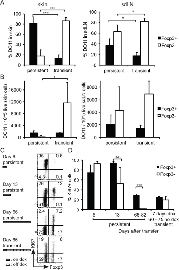
Persistent Ag expression leads to a specific loss of Teff cells in the periphery and relative preservation of Treg. (A) DO11/Rag−/− cells were adoptively transferred to K5/TGO/TCRα−/− and recipient mice were either fed with dox for 7 days and then switched back to regular feed (transient expression) or kept on dox feed for the entire time of the experiment (persistent expression). Single cell suspensions of skin (left) and sdLNs (right) were prepared between days 60–82 post transfer and analyzed by FACS. Percentage of Foxp3+ and Foxp3− T cells within the skin-infiltrating DO11 T cell population. Data pooled from three independent experiments. (Total number of mice analyzed n = 3–5/condition; error bars show SD) (B) As (A), showing the absolute number of Foxp3+ and Foxp3− DO11 T cells/105 live cells in the skin (left) or sdLN (right). (C) sdLN cells were analyzed for expression of Foxp3 and Ki67 on indicated days after DO11 transfer. Data are gated on live donor CD4+, CD90.1+ cells. (D) The percentage of Ki67-expressing LN DO11 cells within the Fopx3+ and Foxp3− population, respectively, was calculated. (Error bars show SD; n = 2–3/condition and time point.)