
FIGURE 3.
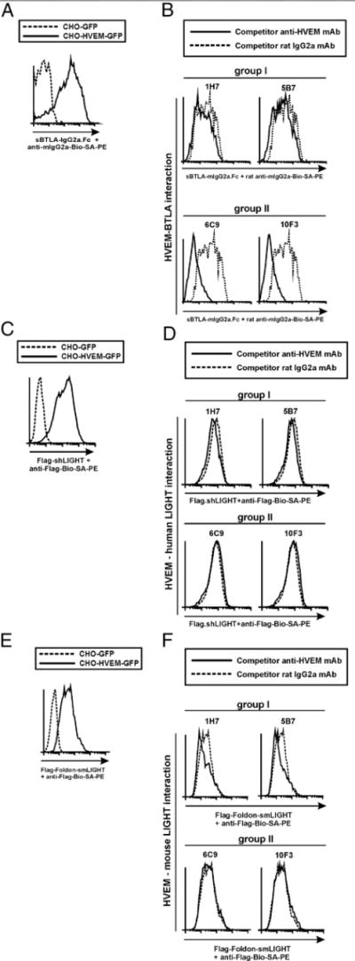
Group II anti-HVEM mAbs exhibit antagonist activity and abrogate HVEM/BTLA interaction without affecting HVEM/LIGHT binding. (A) The specificity and binding affinity of recombinant sBTLA-Ig fusion protein to membrane-bound HVEM-GFP stably transfected CHO cells or control CHO-GFP cells are shown on gated GFP-positive cells. Dashed line corresponds to binding of sBTLA-Ig to CHO-GFP control-transfected cells, and solid line depicts the binding of sBTLA-Ig to HVEM-GFP–transfected cells. (B) A total of 2.5 × 10⁵ stably HVEM-GFP–transfected CHO cells was incubated with 10 μg/ml either rat IgG2a competitor control (dashed lines) or anti-HVEM mAbs (solid lines) following group I (upper panel) or group II (lower panel) for 30 min at room temperature. Then, 10 μg/ml sBTLA-Ig was added to the cells and incubated at 37°C for 2 h. The reaction was washed and further incubated with biotinylated rat anti-mouse IgG2a mAb, and the staining was finally developed with SA-PE. (C) The specificity of Flag-shLIGHT binding to HVEM-GFP–transfected cells (solid line) is shown. Dashed line displays the background binding of Flag-shLIGHT to control GFP-transfected cells. (D) Stably transfected HVEM-GFP cells were incubated with 10 μg/ml either rat IgG2a competitor Ab control (dashed lines) or anti-HVEM mAbs (solid lines) following group I (D, upper panel) or group II (D, lower panel) for 30 min at room temperature. Then, 10 μg/ml Flag-shLIGHT was added to the reaction and incubated at 37°C for 2 h. Biotinylated anti-Flag mAb followed by SA-PE was used to develop the immunological reactions. (E) The specificity of Flag-Foldon-smLIGHT binding to HVEM-GFP–transfected cells (solid line) is depicted. Dotted line represents the background binding of Flag-Foldon-smLIGHT to control GFP-transfected cells. (F) Stably transfected HVEM-GFP cells were incubated with 10 μg/ml either rat IgG2a competitor Ab control (dashed lines) or anti-HVEM mAbs (solid lines) following group I (F, upper panel) or group II (F, lower panel) for 30 min at room temperature. Then, 10 μg/ml Flag-Foldon smLIGHT was added to the reaction and incubated at 37°C for 2 h. Biotinylated anti-Flag mAb followed by SA-PE was used to develop the immunological reactions. A cartoon of the competition assay setup is shown. One representative experiment of three with identical results is shown.