
FIGURE 4.
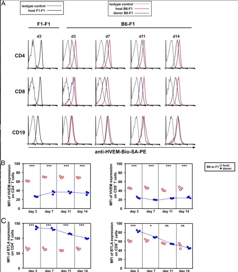
Reciprocal regulation of HVEM and BTLA expression is dependent on alloantigen recognition by donor T cells during the course of GvHR. (A) A total of 70 × 106 of B6 splenocytes was adoptively transferred into F1 recipients (B6-F1), and the time course of HVEM expression was monitored at days 3, 7, 11, and 14 after GvHR induction on host and donor CD4+ T cells, CD8+ T cells, and CD19+ cells of spleen. Syngeneic adoptive transfer of F1 splenocytes to F1 recipients (F1-F1) served as control group to determine the basal level of expression of these molecules at resting state under noninflammatory conditions. Black solid lines represent basal expression of HVEM on host cells of F1 mice receiving syngeneic F1 splenocytes; black dashed lines display isotype-matched control. Allogeneic adoptive transfer of B6 splenocytes to F1 recipients allowed the monitoring of surface expression of HVEM receptor on host (red solid lines) and donor (blue solid lines) lymphocytes during the course of GvHR. Black dashed lines indicate isotype-matched control. Biotinylated anti-HVEM mAb (10F3) followed by SA-PE was used to develop the reaction. The mean fluorescence intensity (MFI) of HVEM (B) and BTLA (C) expression on CD4 and CD8 T cells was calculated at different time points after the adoptive transfer of allogeneic splenocytes to F1 recipient mice. Red open circles and blue closed circles depict values of MFI of host and donor T cells, respectively. At each time point, MFI of HVEM and BTLA expression on donor and host CD4 and CD8 T cells was compared, and the statistical significant differences are indicated inside the plots (*p < 0.05, **p < 0.005, ***p < 0.0005; ns, nonsignificant). Blue dotted line highlights the expression trend of HVEM and BTLA on donor CD4 and CD8 T cells.