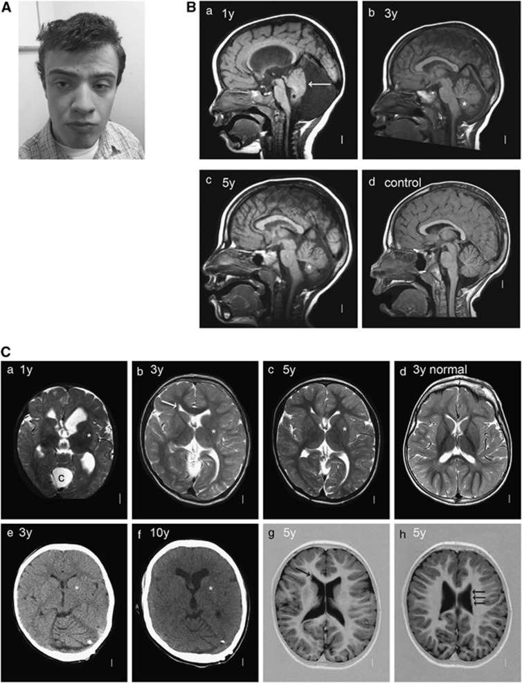
Figure 2.
(A) Photograph of affected individual V.1 at 16 years of age shows a long face, broad forehead, deep-set eyes, large mouth with full lips and prominent jaw. (B) Midline sagittal T1-weighted MRI scans of patient V.1 at ages 1 (a), 3 (b) and 5 (c) years compared with a normal control (d). The first scan (a) shows a very large, fluid-filled posterior fossa and flattening of the posterior vermis (white arrow). The inferior portion of the posterior vermis is abnormally small in all three scans (asterisks in a–c). (C) Serial brain imaging studies in patient V.1 including axial MRI T2-weighted MRI images at 1 (a), 3 (b) and 5 (c) years, and in a normal control (d) for comparison; CT scans at 3 (e) and 10 (f) years of age; and inverted T2-weighted images at 5 years (g and h). The first scan (a) shows moderately enlarged 3rd and lateral ventricles and a large posterior fossa cyst (c). All CT and MRI images show normal basal ganglia (asterisks placed over the left putamen in a–c and e, f). Multiple periventricular nodular heterotopia are seen along the right frontal horn (arrow in b and g) and left mid-body (h) of the lateral ventricles.

