
Fig. 6.
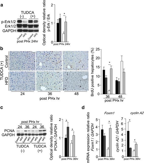
TUDCA pretreatment improved delayed liver regeneration in simple fatty liver. a The activation of Erk1/2 at 24 h after PHx with or without TUDCA pretreatment evaluated by Western blotting. b BrdU immunohistochemical staining (original magnification ×200) with TUDCA pretreatment, and BrdU labeling index at 36 and 48 h after PHx with or without TUDCA pretreatment. c The expression of PCNA protein at 24 and 36 h after PHx with or without TUDCA pretreatment detected by Western blotting, densitometric analysis was performed at 36 h after PHx. d The mRNA levels of Foxm1 and cyclin A2 at 36 h after PHx with or without TUDCA pretreatment measured by real-time RT-PCR (black squares Control, dark gray squares Control with TUDCA, white squares HFD, light gray squares HFD with TUDCA, n = 4–6; mean ± SE, *p < 0.05 mice with TUDCA pretreatment vs. mice without TUDCA pretreatment fed same diet in each time point by ANOVA and Wilcoxon test)