


Figure 1. Morphology of germinal matrix.
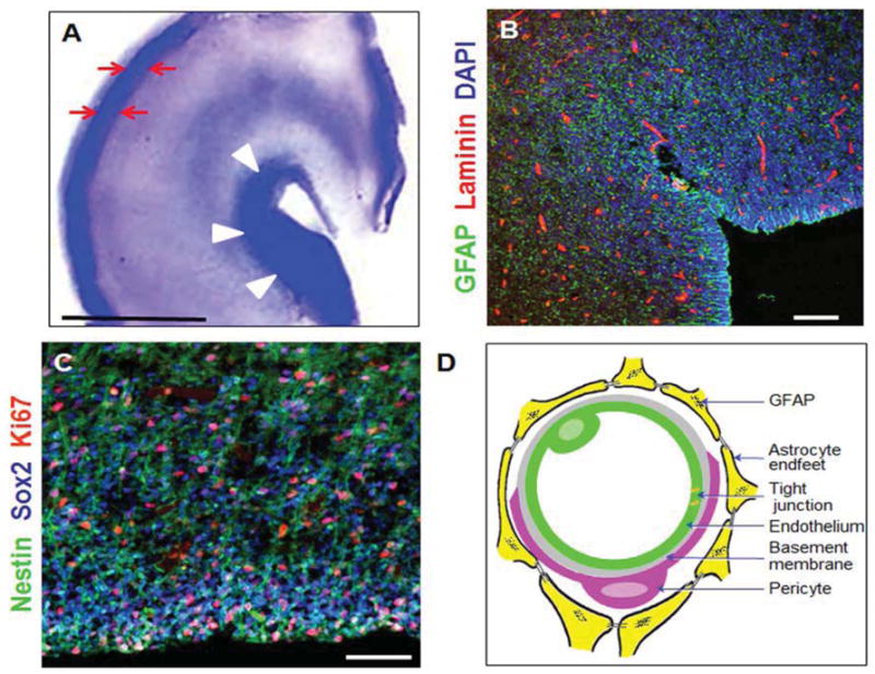
A) Representative cresyl violet staining of coronal section of the right-sided cerebral hemisphere of a 23 week preterm infant. Note cortical plate (arrows) and germinal matrix (arrow heads). Germinal matrix (violet staining) surrounds the whole ventricle, but is most conspicuous at the head of caudate nucleus. V, ventricle. Scale bar, 0.5 cm. B) Representative immunofluorescence of cryosection from germinal matrix of a 23 week premature infant labeled with DAPI (blue), GFAP (green), and laminin (vascular marker, red). Note germinal matrix is highly vascular (vascular endothelium in red) and enriched with GFAP (+) glial cells (green). C) Coronal brain section was double labeled with nestin (progenitor cells, green), Sox2 (radial glia, blue), and Ki67 (red, proliferation marker). Note nestin and Sox2 positive cells are abundant in the germinal matrix. Scale bar; 100 (B) and 50 μm (C). D) Schematic drawing of the blood brain barrier in cross section showing endothelium, endothelial tight junction, basal lamina, pericyte, and astrocyte endfeet.