
Figure 2.
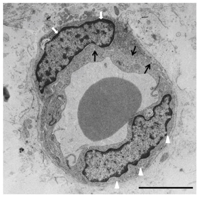
Electron micrograph showing endothelium (arrow heads) and pericyte (white arrows) separated by basal laminia (black arrows) in the white matter of 3 day old preterm rabbit pup (E29). Note pericyte wraps around the endothelium and is outer to the basal lamina. Scale bar, 4 μm.