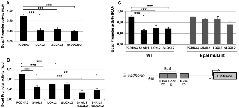
Fig. 2. E-cadherin is transcriptionally repressed by LOXL2.
(A) The activity of the E-cadherin promoter in HEK293T cells was measured in the presence of the indicated LOXL2 variants (50 ng). (B) E-cadherin promoter activity in HEK293T cells was measured in the presence of the indicated LOXL2 forms (50 ng) and in the absence or presence of Snail1 (50 ng). The effect of Snail1 (50 ng) in the absence of LOXL2 and ΔLOXL2 was also tested. (C) Activity of the wild-type (left) and mutant E-cadherin promoter (Epal mutant) (right) in HEK293T cells was measured in the presence the indicated factors (50 ng). Schematic representation of the proximal mouse E-cadherin promoter is shown at the bottom. In all cases, the activity was determined as relative luciferase units (RLU) and normalized to the activity detected in the presence of control pcDNA3 vector. Results represent the mean ± s.e.m. of at least three independent experiments performed in triplicate samples (*p<0.05, **p<0.005, ***p<0.001).

