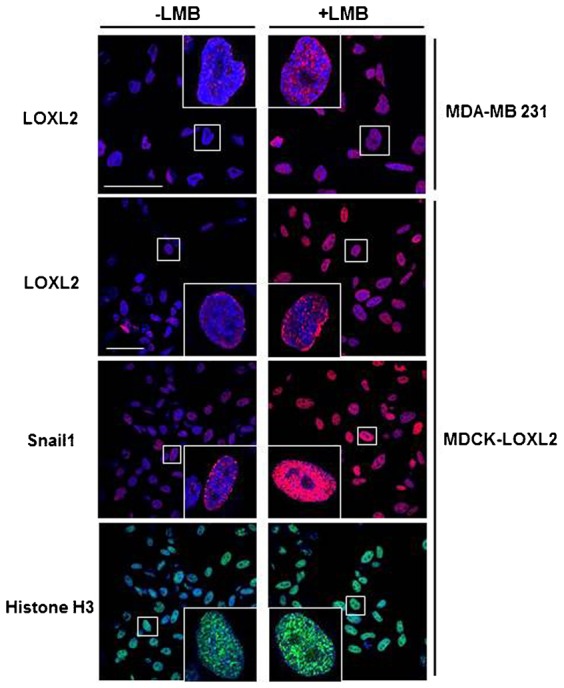
Fig. 4. Translocation of LOXL2 into the nucleus.

Accumulation of LOXL2 within the nucleus in MDA-MB231 or MDCK-LOXL2 cells was analyzed by confocal immunofluorescence in the absence or presence of Leptomycin B (LMB; 5 ng/ml, 16 h). Distribution of Snail1 and histone H3 were also included as control. Images show only the overlap between red (LOXL2 and Snail1) or green signals (Histone H3) and DAPI staining. Scale bars: 50 µm.
