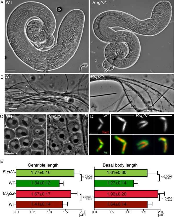
Fig. 3. Characterisation of Bug22 testes.
(A) Pictures from adult WT (left) and Bug22 testes (right) showing regular testes morphology. Scale bar: 100 µm. (B) High magnification pictures of WT (left) and Bug22 (right) spermatids showing the presence of thicker sperm tails in the mutant. Scale bar: 10 µm. (C) Phase contrast image showing that Bug22 (right) onion stage spermatids are indistinguishable from WT (left). Every post-meiotic round spermatid contains one nucleus (white circle) adjacent to one nebenkern (black circle), both structures having approximately the same size. Onion stages from at least 20 males were analysed. Scale bar: 10 µm. (D) Images of WT and Bug22 dividing primary spermatocyte centrioles expressing RFP-PACT (top panel and shown in red in the merged panel) and Asl (shown in green in the merged panel). Scale bar: 2 µm. (E) Graphs showing measurements of centriole (left) and basal body (right) lengths in WT and Bug22. Measurements were based on the fluorescence signal of the transgenic protein RFP-PACT (red bars) and Asl (green bars). Mean values of length (±) represent the standard deviation from more than 50 centrioles from meiosis I or II spermatocytes (left) and elongated spermatids (right). Student's t-tests were performed to assess statistical differences.

