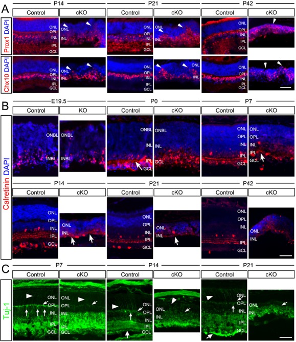
Fig. 2. Aberrant retina lamination and loss of plexiform layers caused by Top2b deletion.

(A) Retina sections prepared from P14, P21 and P42 control and Top2b cKO mice were stained with DAPI and co-stained with either Prox1 or Chx10. Prox1+ and Chx10+ cells were detected in the ONL (arrowheads) in Top2b cKO retinas. (B) Co-staining of E19.5-P42 control and KO/cKO retina sections with DAPI and Calretinin, which labels neurofilaments in the IPL. In cKO retinas, only a few filament-like structures were found at P7, P14 and P21, while in control samples the strata structure of neurofilaments appeared as early as P0 (arrows). (C) Co-staining with Tuj-1in P7, P14 and P21 control and cKO retina sections. Tuj-1 stained neurofilaments of the OPL (arrows) and processes of photoreceptors (arrowheads), which were largely missing in cKO samples. GCL, ganglion cell layer; INL, inner nuclear layer; IPL, inner plexiform layer; ONL, outer nuclear layer; OPL, outer plexiform layer. Scale bars: 50 µm.
