
Fig. 3. Top2b deficiency leads to delayed embryonic development and decreased ganglion and horizontal cells.
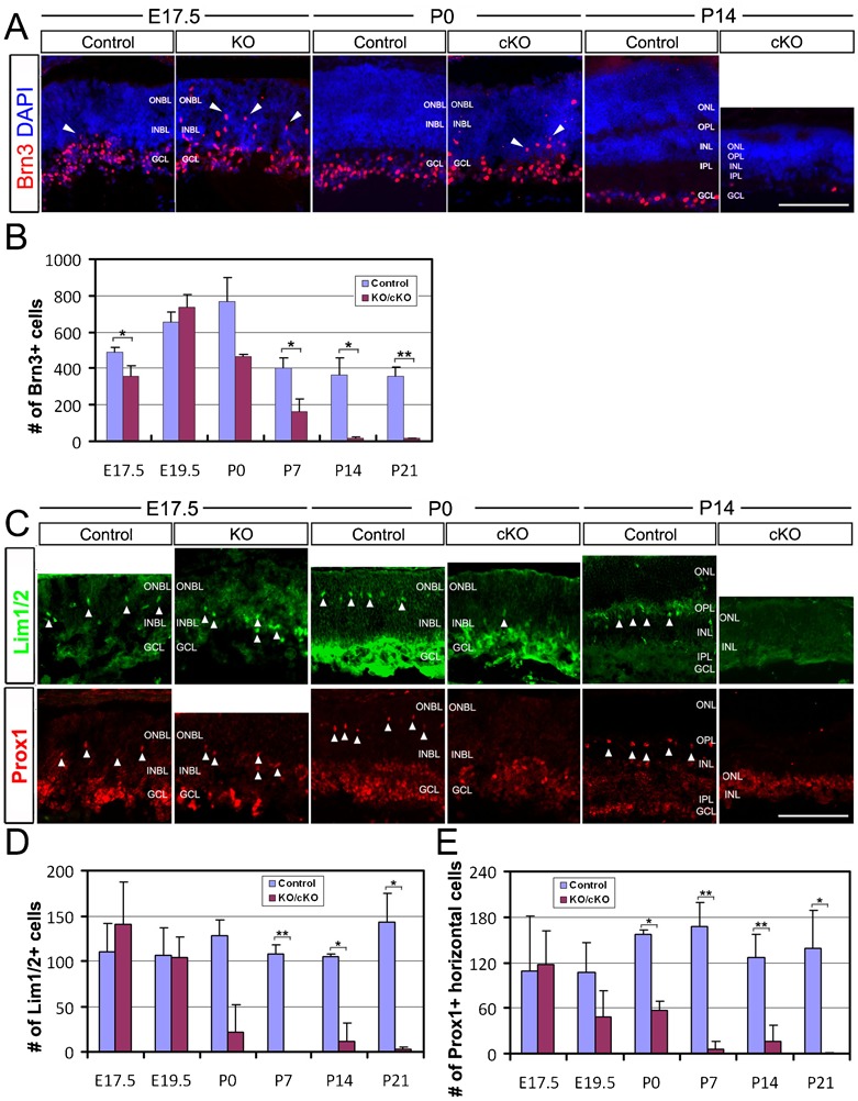
(A) Retina sections of E17.5 control and KO embryos, and P0 and P14 control and cKO mice were co-stained with Brn3 and DAPI. Brn3+ cells were all located in the GCL in the control retinas, but they were more widely distributed in the INBL and ONBL (arrowheads) in the KO/cKO retinas. (B) Quantification of Brn3+ cells. A significant reduction in the number of Brn3+ cells in postnatal stages starting from P7 was observed. (C) Retina sections of E17.5 control and KO embryos and P0 and P14 control and cKO mice were co-stained with Lim1/2 and DAPI or Prox1 and DAPI. Lim1/2+ and Prox1+ horizontal cells were well-spaced and located in the horizontal cell layer (arrowheads) in the control retinas; while in the KO/cKO retinas, the majority of these cells remain in the INBL (arrowheads) at E17.5 and P0. By P14, no Lim1/2+ or Prox1+ cells were detected in cKO samples. (D) Quantification of Lim1/2+ cells. (E) Quantification of Prox1+ horizontal cells. Error bars are s.d. (n = 3 except n = 2 for P0). Student's t-test, *p<0.05, **p<0.01. INBL, inner neuroblastic layer; ONBL, outer neuroblastic layer; GCL, ganglion cell layer; INL, inner nuclear layer; IPL, inner plexiform layer; ONL, outer nuclear layer; OPL, outer plexiform layer. Scale bars: 100 µm.