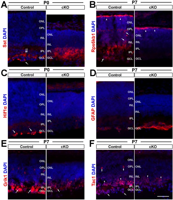
Fig. 7. Top2b deletion affects the expression of genes involved in neural cell survival and neurite growth.

Control and cKO retina sections of P0 and P7 mice were stained with antibodies against six proteins with their respective mRNA differentially expressed in cKO retinas. (A) Sst staining in cKO sample was increased but lost the specific immunoactivity in the GCL (double arrow) and ganglion cells (arrow) in the control retinas. (B) The Rps6kb1 protein was expressed in the outer segments of photoreceptors (arrowheads), ganglion cells, amacrine cells (arrows) and the IPL in the control retinas. However, its expression was only seen in the outer segments (arrowheads) in cKO retinas. (C) Hif1α was found in the cytoplasm of marginal ganglion cells in the controls (arrows), but was not detectable in the cKO samples. (D) Increased GFAP expression was found in GCL of P7 cKO retinas. (E) Grik1 was strongly stained in the IPL of the control retina, but reduced in the cKO retina (arrows). In addition, Grik1 staining in horizontal cells was missing. (F) Tac1 was found in horizontal cells (vertical arrows), amacrine cells (arrowheads) and ganglion cells (diagonal arrows) in the control retina. In the cKO retina, the signal was only found in amacrine cells with a decreased intensity. GCL, ganglion cell layer; INL, inner nuclear layer; IPL, inner plexiform layer; ONL, outer nuclear layer; OPL, outer plexiform layer. Scale bar: 50 µm.
