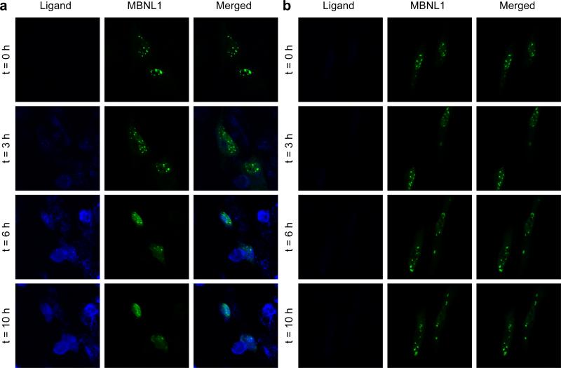
Figure 7.
Live cell microscopy demonstrates a direct evidence for MBNL1 foci dispersion with 9. a) Live DM1 cell model are treated with 9 (50 μM) at t = 0, immediately after the first image is taken. Fluorescence of 9, confirms its penetration to the nucleus. MBNL1 nuclear foci are gradually dispersing over time in two cells. b) Two live cell show stability of foci in DM1 cell model, in the absence of 9, over the period of 10 h. Each box shows 120 μM X 120 μM.

