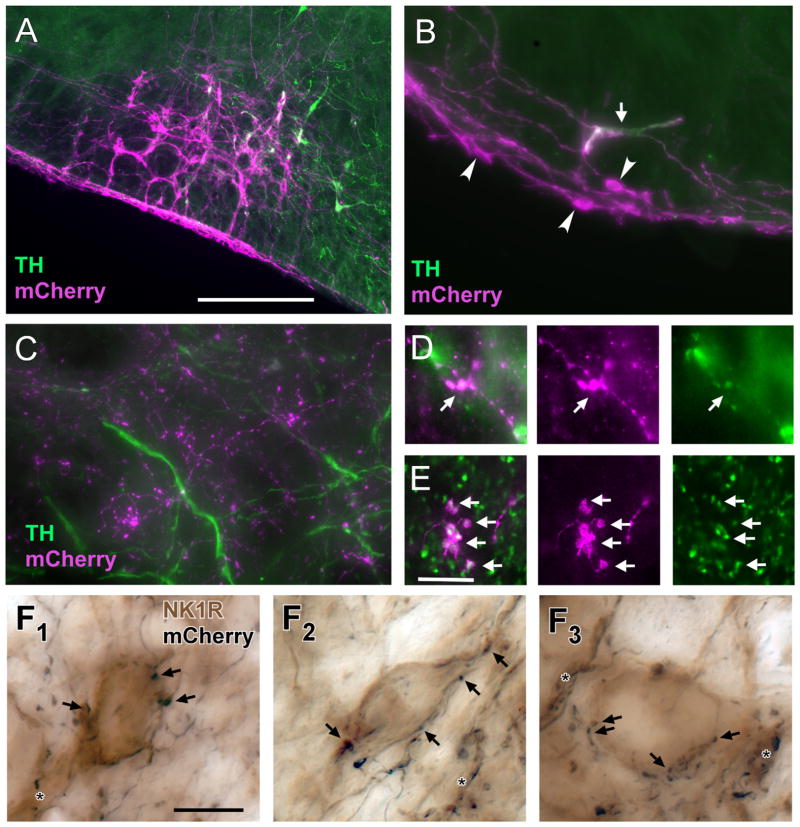
Figure 5.
Terminals of RTN-Phox2b neurons make close appositions with NK1R-ir neurons in the pre-Bötzinger complex. A: Example of mCherry-ir neurons within the RTN region 3 weeks after injection of PRSX8 ChR2-mCherry lentivirus into rats pretreated with anti-DβH-saporin to reduce the number of spinally projecting TH neurons. The vast majority of the transfected neurons are not TH-ir (TH immunofluorescence in green; ChR2-expressing TH neurons in white). B: Higher magnification example showing a single TH-ir neuron (arrow) and multiple TH-negative neurons (arrowheads) that contain mCherry (magenta). The depicted region is below the facial motor nucleus. The TH-negative neurons are located within the marginal layer of the RTN. C: The mCherry-ir terminals (magenta) located within the pre-Bötzinger complex do not contain TH immunoreactivity (green). D: mCherry-ir terminals (magenta) located within the raphe pallidus are TH-ir (TH in green, double-labeled terminals appear white). E: A large fraction of the mCherry-ir (magenta) terminals located within the Pre-Bötzinger complex are positive for VGLUT2 (green, double-labeled terminals appear white). F1–F3: Examples of close appositions between mCherry-ir varicosities (nickel-enhanced DAB reaction, black) and NK1R-ir somata or dendrites within the pre-Bötzinger complex (light brown). Some of the close appositions (putative synapses) are indicated by arrows. Close appositions on dendrites are indicated by the asterisks. Scale bar = 250 μm in A (applies to A–C); 10 μm in E (applies to D, E); 10 μm in F1 (applies to F1–F3).

