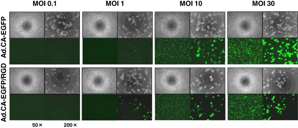
Figure 3.

AGTEs of adenoviral vectors with wild or modified fiber. KYM-1 cells were infected with either Ad.CA-EGFP or Ad.CA-EGFP/RGD, which have wild-type or modified fiber (RGD-peptide added to the fiber knob), respectively, at an MOI of 0.1, 1, 10, or 30, and then observed under phase-contrast and fluorescence microscopy 48 h later. Representative phase-contrast (upper) and fluorescence (lower) images are shown at 50× (left) and 200× (right) magnification. There was no apparent difference in AGTEs between the two groups.
