Figure 5.

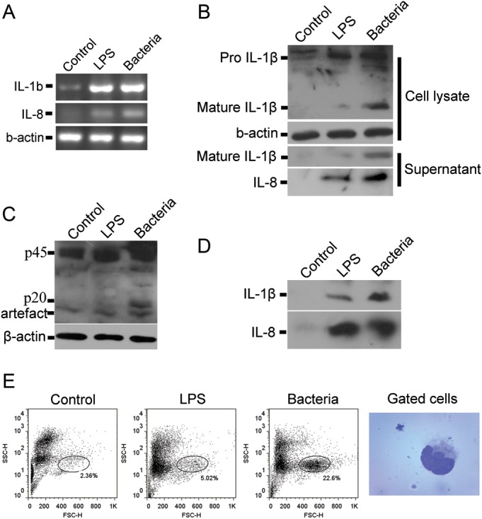
Lipopolysaccharide (LPS) and bacteria induced pro-inflammatory caspase activation and pro-inflammatory cytokine secretion in vitro and in vivo. (A) Splenocytes were isolated from B. maxima spleen and stimulated with LPS (8 μg/ml) or bacteria (3 × 10⁸cfu/ml) for 2 h. The cells were collected for RT-PCR of IL-1βand IL-8. Either LPS or bacteria can upregulate the mRNA level of IL-1β and IL-8. (B) Splenocytes were incubated with LPS or bacteria. The cells and the supernatants were collected for western blotting of IL-1β and IL-8. (C) Splenocytes stimulated with LPS or bacteria were lysed for the detection of caspase-1 activation by western blotting. The band of ∼20 kDa represents the presence of activated caspase-1. (D) Bombina maxima weighted 25 ± 5 g were injected intraperitoneally with LPS (50 mg/kg in 0.1 ml PBS) or bacteria (3 × 10⁸cfu in 0.1 ml PBS, Staphylococcus aureus, ATCC25923). Two hours after injection, peritoneum exudates were harvested, IL-1β and IL-8 were detected by western blot. (E) Two hours after injection, peritoneal cells were harvested and analysed by flow cytometry. The mostly changed subset of cells was stained and photographed.
