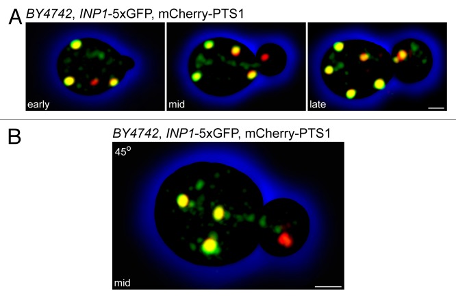
Figure 1. Inp1p is present in small vesicles that are distinct from the peroxisomal compartment. Wild-type yeast cells expressing Inp1p-5 × GFP and mCherry-PTS1 were visualized by confocal fluorescence microscopy. Images were acquired as 3D z-stacks and flattened into maximum intensity projections. (A) Time course representing early, mid, and late stages of peroxisome inheritance in a single cell. Bar, 1 μm. (B) A budding cell at the mid-stage of peroxisome inheritance. Individual z-stacks were combined and displayed at a 45° angle to enhance the 3D effect. Bar, 1 μm.

An official website of the United States government
Here's how you know
Official websites use .gov
A
.gov website belongs to an official
government organization in the United States.
Secure .gov websites use HTTPS
A lock (
) or https:// means you've safely
connected to the .gov website. Share sensitive
information only on official, secure websites.
