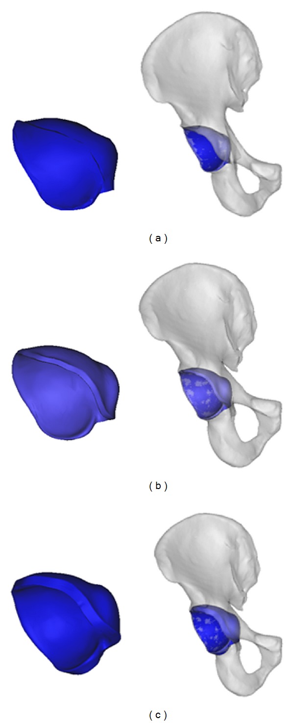
Figure 1.

The acetabular model was stimulated from the pelvic model and “shelled” for 2.95 mm and 6 mm, respectively. (a) The acetabular model was extracted from the pelvic model; (b) the acetabular model was “shelled” for 2.95 mm and assembled with pelvic model; (c) the acetabular model was “shelled” for 6 mm and assembled with pelvic model.
