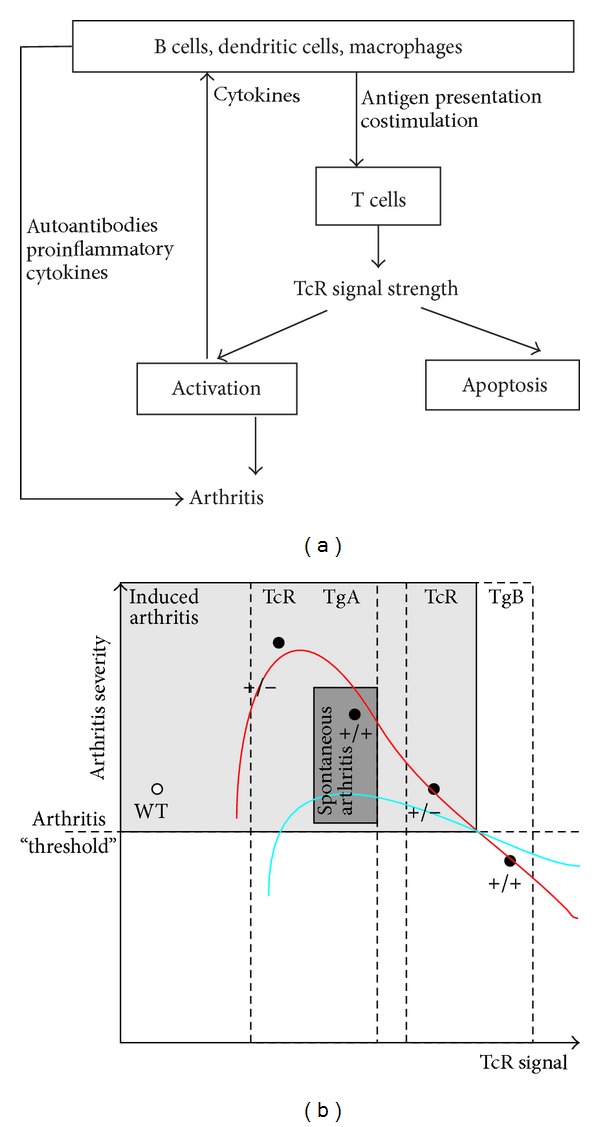
Figure 3.

The balance of T cell activation and apoptosis regulates the development of autoimmune arthritis in PGIA. (a) The interaction between APCs and T cells determines T cell activation. TCR signal strength is regulated by costimulation and TCR expression. When the activation signal is optimal, T cell activation will lead to the autoimmune attack of joints. (b) Relation between the strength of the TCR signal and arthritis severity in the PG-specific TcR-Tg mice. The two mouse colonies (TCR-TgA and TCR-TgB) were described in [13]. The use of homo- (+/+) or heterozygous (+/−) TCR-Tg mice allowed us to further refine our TCR signal strength studies in PGIA. Activation (red line) or apoptosis (blue line) of T cells is regulated by TCR signal strength. When the activation exceeds apoptosis, the autoreactive cells accumulate and arthritis develops (grey area). When apoptosis overrides activation the autoreactive T cells are eliminated and arthritis does not develop. Spontaneous arthritis (dark grey zone) was only observed in homozygous TCR-TgA mice [14]; thus, at least from the T cell signaling side, a very narrow “window” exists when T cell activation is optimal and is over the apoptosis.
