Figure 1.

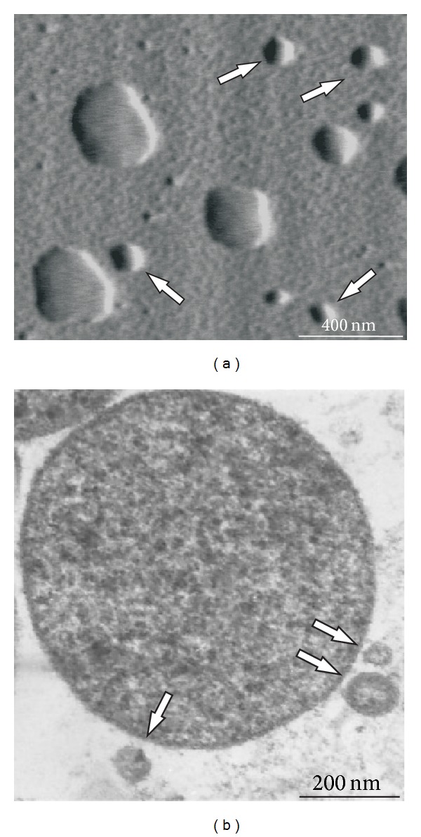
Atomic force microscopy (a) and transmission electron microscopy (b) images of Acholeplasma laidlawii PG8 cells and extracellular vesicles (indicated by arrows).

Atomic force microscopy (a) and transmission electron microscopy (b) images of Acholeplasma laidlawii PG8 cells and extracellular vesicles (indicated by arrows).