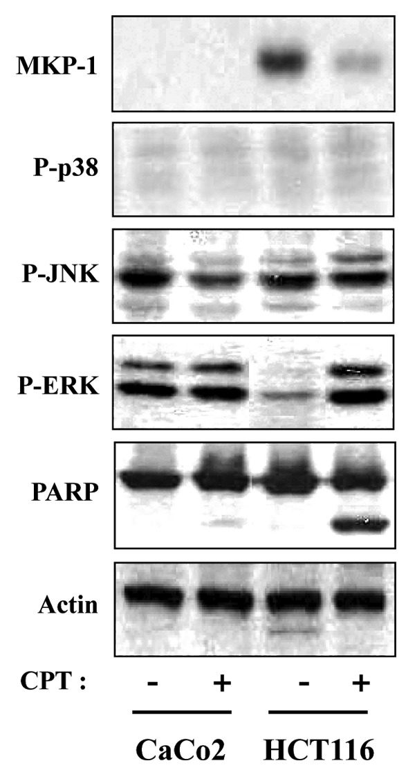
Figure 2. CPT-induced apoptosis in HCT116 cells, but not CaCo2 cells, is associated with decreased expression of MKP1. HCT116 and CaCo2 cells were treated with 500 nM and 1000 nM CPT for 24 h respectively, and total cell extracts were prepared and subjected to western blot analysis using anti-MKP1 (MKP1), anti-phospho-p38 (P-p38), anti-phospho-JNKs (P-JNK), anti-phospho-ERKs (P-ERK), and anti-PARP antibodies. Variations in protein loading among samples were controlled by monitoring actin levels.
