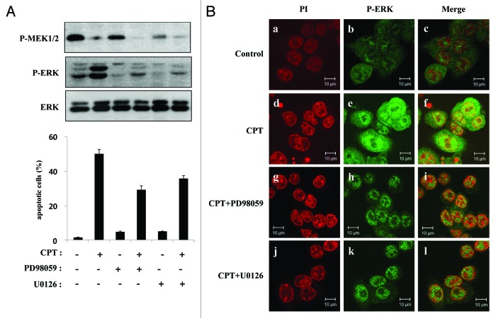
Figure 6. MEK inhibitors attenuate CPT-induced apoptosis and ERK activation. (A) HCT116 cells were exposed to 500 nM CPT. One hour after treatment, the cells were treated with PD98059 (30 µM) or U0126 (10 µM) in the presence or absence of CPT, as indicated. After an additional 24 h, total cell extracts from HCT116 cells were analyzed by western blotting using primary antibodies to phospho-MEK1/2 (P-MEK1/2), phospho-ERKs (P-ERK), and total ERKs (ERK). Apoptosis levels were determined using PI staining and flow cytometry under the same conditions as above. (B) HCT116 cells were treated with CPT (500 nM), PD989059 (30 µM), or U0126 (10 µM), as indicated, using the same conditions as above. After 24 h, cells were fixed and immunostained for phosphorylated ERKs using a FITC-conjugated secondary antibody and counterstained with PI. (a, d, g, and j) Staining for PI (red). (b, e, h and k) Localization of phosphorylated ERKs (green). (c, f, I, and l) Merged images of phosphorylated ERKs and PI.

An official website of the United States government
Here's how you know
Official websites use .gov
A
.gov website belongs to an official
government organization in the United States.
Secure .gov websites use HTTPS
A lock (
) or https:// means you've safely
connected to the .gov website. Share sensitive
information only on official, secure websites.
