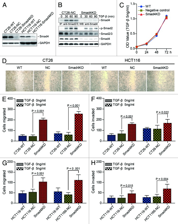
Figure 2. Smad4 knockdown promotes the migration and invasion of CT26 and HCT116 cells in vitro. (A) CT26-Smad4KD, HCT116-Smad4KD, and their wild type, negative control cells were subjected to western blot analysis with the indicated antibodies. (B) CT26-NC and CT26-Smad4KD cells were treated with TGF-β1 (10 ng/ml) for the indicated time and the lysates were immunoprecipitated by Smad2/3 antibody followed by western blotting with Smad4 antibody (top panel). Total cell lysates were subjected to western blot analyses with the indicated antibodies (bottom panel). GAPDH served as loading control. (C) CT26-WT, CT26-NC, and Smad4KD cells were treated with or without TGF-β (5 ng/ml) for 72 h. Cells were subjected to CCK-8 assay. (D) CT26-WT, CT26-NC, CT26-Smad4KD, HCT116-WT, HCT116-NC, and HCT116-Smad4KD cells were treated with TGF-β (5 ng/ml), and subjected to wound healing assay. (E and F) CT26-WT, CT26-NC, and Smad4KD cells were treated with or without TGF-β (5 ng/ml), and subjected to transwell migration and invasion assay. The data were presented as the mean ± SD (n = 6). (G and H) HCT116-WT, HCT116-NC, and HCT116-Smad4KD cells were treated with or without TGF-β (5 ng/ml), and subjected to transwell migration and invasion assay. The data were presented as the mean ± SD (n = 6).

An official website of the United States government
Here's how you know
Official websites use .gov
A
.gov website belongs to an official
government organization in the United States.
Secure .gov websites use HTTPS
A lock (
) or https:// means you've safely
connected to the .gov website. Share sensitive
information only on official, secure websites.
