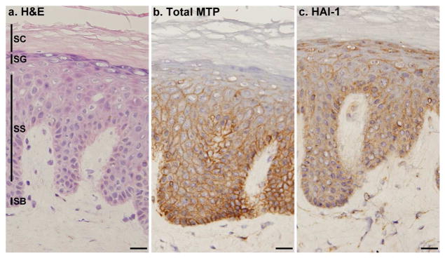
Figure 2. Cellular localization of matriptase and HAI-1 in normal human skin.

Frozen sections from normal human skin were stained with hematoxylin and eosin (a. H&E) or immunostained with mAb M32 for total matriptase (b. Total MTP) or mAb M19 for HAI-1 (c. HAI-1). Nuclei of cells in all three panels were counterstained blue with hematoxylin. The epidermal layers are indicated by SB for the stratum basale, SS for the stratum spinosum, SG for the stratum granulosum and SC for the stratum corneum. Scale bar: 25 μm.
